News
No news have been found.
-
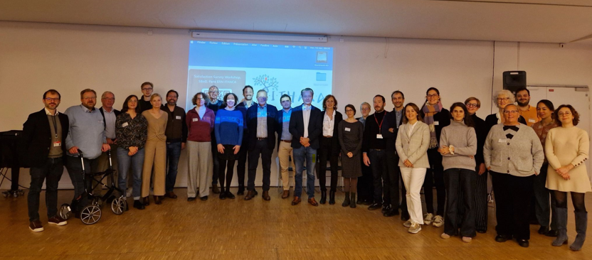
Workshop, SBoD Work Group – ERN ITHACA, eUROGEN, ERKnet – 14-15 November 2024 – Paris APHP
This two-day workshop, held from November 14-15, offers an in-depth exploration of current research and advances in genetic and perinatal…
-
SBoD webinar on the new classification of dysraphisms
This is a series of webinars on spinal dysraphism organised jointly by ERN eUROGEN, OMNI-NET Ukraine and the International Federation…
-
SBoD: New article on prenatal diagnosis of open dysraphism
The European SBoD (Spina Bifida and other Dysraphisms) working group, co-chaired by Professor Jean-Marie Jouannic, has published a new article…
Our publications
Blondeau, Alicia; Laraqui, Samia; Mendes, Goncalo; Hascoet, Juliette; Haudebert, Camille; Brochard, Charlène; Richard, Claire; Voiry, Caroline; Siproudhis, Laurent; Timoh, Krystel Nyangoh; Manunta, Andrea; Samson, Emmanuelle; Peyronnet, Benoit
In: Fr J Urol, vol. 35, no. 13-14, pp. 102938, 2025, ISSN: 2950-3930.
@article{pmid40714359,

title = {Synthetic mid-urethral slings in female spina bifida patients with stress urinary incontinence: Is it worth it?},

author = {Alicia Blondeau and Samia Laraqui and Goncalo Mendes and Juliette Hascoet and Camille Haudebert and Charlène Brochard and Claire Richard and Caroline Voiry and Laurent Siproudhis and Krystel Nyangoh Timoh and Andrea Manunta and Emmanuelle Samson and Benoit Peyronnet},

doi = {10.1016/j.fjurol.2025.102938},

issn = {2950-3930},

year  = {2025},

date = {2025-12-01},

journal = {Fr J Urol},

volume = {35},

number = {13-14},

pages = {102938},

abstract = {INTRODUCTION: The aim of this study was to report the outcomes of synthetic mid-urethral slings (MUS) in female patients with spina bifida and stress urinary incontinence (SUI).nnMETHODS: All female patients with spina bifida who were seen at a national referral center between 2007 and 2021 and who had a history of MUS for SUI were included in a retrospective study. The primary outcome of interest was the continence status at 1year as per patients' subjective perception categorized as complete continence, improved continence, unchanged or worsened SUI.nnRESULTS: Out of 339 female spina bifida patients; 11 patients had undergone a MUS insertion and were included for analysis (3.2%). The median age was 30years (range: 19-52years). There were four postoperative complications (36.4%). The three patients fully continent at three months were still fully continent at one year (27.3%) while three had improved continence (27.3%) and five had unchanged continence (45.5%). After a median follow-up of 102months, only one patient was still fully continent (9.1%) and three patients had still improved continence (27.3%). Five patients (45.5%) underwent a subsequent anti-incontinence procedure. The only adverse predictor of improved/complete continence at 1year was a sacral neurological level (OR=0.05; p=0.03).nnCONCLUSION: MUS in women with spina bifida and SUI appears to be safe but less effective than in the non-neurogenic population which may be explained by different underlying pathophysiological mechanisms. These findings question the relevance of this treatment option in these complex population, especially in the current mesh controversy era.},

keywords = {},

pubstate = {published},

tppubtype = {article}

}
Battesti, Guillaume; Chomienne, Lucas; Severac, François; Finoco, Mickael; Ferrero, Emmanuelle; Khalifé, Marc; Odent, Thierry; de Charnace, Edouard; Pesenti, Sebastien; Prost, Solène; Blondel, Benjamin; Solla, Federico; Ould-Slimane, Mourad; Illhareborde, Brice; Charles, Yann Philippe
2025, ISSN: 1432-0932.
@misc{pmid41369755,

title = {Correction: Global and segmental thoracolumbar sagittal alignment in adolescent idiopathic scoliosis compared to normal individuals},

author = {Guillaume Battesti and Lucas Chomienne and François Severac and Mickael Finoco and Emmanuelle Ferrero and Marc Khalifé and Thierry Odent and Edouard de Charnace and Sebastien Pesenti and Solène Prost and Benjamin Blondel and Federico Solla and Mourad Ould-Slimane and Brice Illhareborde and Yann Philippe Charles},

doi = {10.1007/s00586-025-09605-2},

issn = {1432-0932},

year  = {2025},

date = {2025-12-01},

journal = {Eur Spine J},

keywords = {},

pubstate = {published},

tppubtype = {misc}

}
Pelvic organ prolapse is highly prevalent in women with spina bifida
Dubois, Alexandre; Malandain, Briac; Hascoet, Juliette; Haudebert, Camille; Brochard, Charlène; Richard, Claire; Voiry, Caroline; Timoh, Krystel Nyangoh; Manunta, Andrea; Samson, Emmanuelle; Peyronnet, Benoit
In: BJUI Compass, vol. 6, no. 11, pp. e70113, 2025, ISSN: 2688-4526.
@article{pmid41293758,

title = {Pelvic organ prolapse is highly prevalent in women with spina bifida},

author = {Alexandre Dubois and Briac Malandain and Juliette Hascoet and Camille Haudebert and Charlène Brochard and Claire Richard and Caroline Voiry and Krystel Nyangoh Timoh and Andrea Manunta and Emmanuelle Samson and Benoit Peyronnet},

doi = {10.1002/bco2.70113},

issn = {2688-4526},

year  = {2025},

date = {2025-11-01},

journal = {BJUI Compass},

volume = {6},

number = {11},

pages = {e70113},

abstract = {INTRODUCTION: Women with spina bifida often experience neurological impairments leading to pelvic organ dysfunction, including difficulties with bladder and bowel emptying that necessitate frequent Valsalva manoeuvres. These factors, combined with pelvic floor weakness, may increase the risk of pelvic organ prolapse (POP). This study aimed to assess the prevalence of POP in women with spina bifida, identify associated risk factors and evaluate outcomes of surgical management.nnMETHODS: We retrospectively analysed a prospectively maintained database of women with spina bifida seen at a French referral centre from 2007 to 2024. Age under 18 and congenital perineal abnormality were exclusion criteria. The primary outcome was the presence of POP grade 2 or higher (Baden-Walker classification). Secondary outcomes included symptomatic POP requiring surgery, recurrence after surgery, use of vaginal pessaries and related symptoms.nnRESULTS: POP grade ≥2 was present in 14.8% of patients. Women with POP were older (median 44 vs. 31 years; p < 0.0001) and more frequently parous (58.5% vs. 18.3%; p < 0.0001), although 41.5% of POP cases occurred in nulliparous women. Apical prolapse was predominant (64.3%). Among 11 patients who underwent POP surgery, 54.5% experienced recurrence. Multivariate analysis identified parity (OR 5.33; p = 0.005) and lower maximum urethral closure pressure (OR 0.97; p = 0.02) as independent risk factors.nnCONCLUSIONS: POP is highly prevalent in young adult women with spina bifida, including many nulliparous patients. The parity status and a low maximum urethral closure pressure could be associated with an increased risk of POP in this population. High recurrence after surgery highlights the need for information, routine screening and tailored management in this population.},

keywords = {},

pubstate = {published},

tppubtype = {article}

}