
Actualités
-
3ᵉ Journée Scientifique du CRMR Spin@
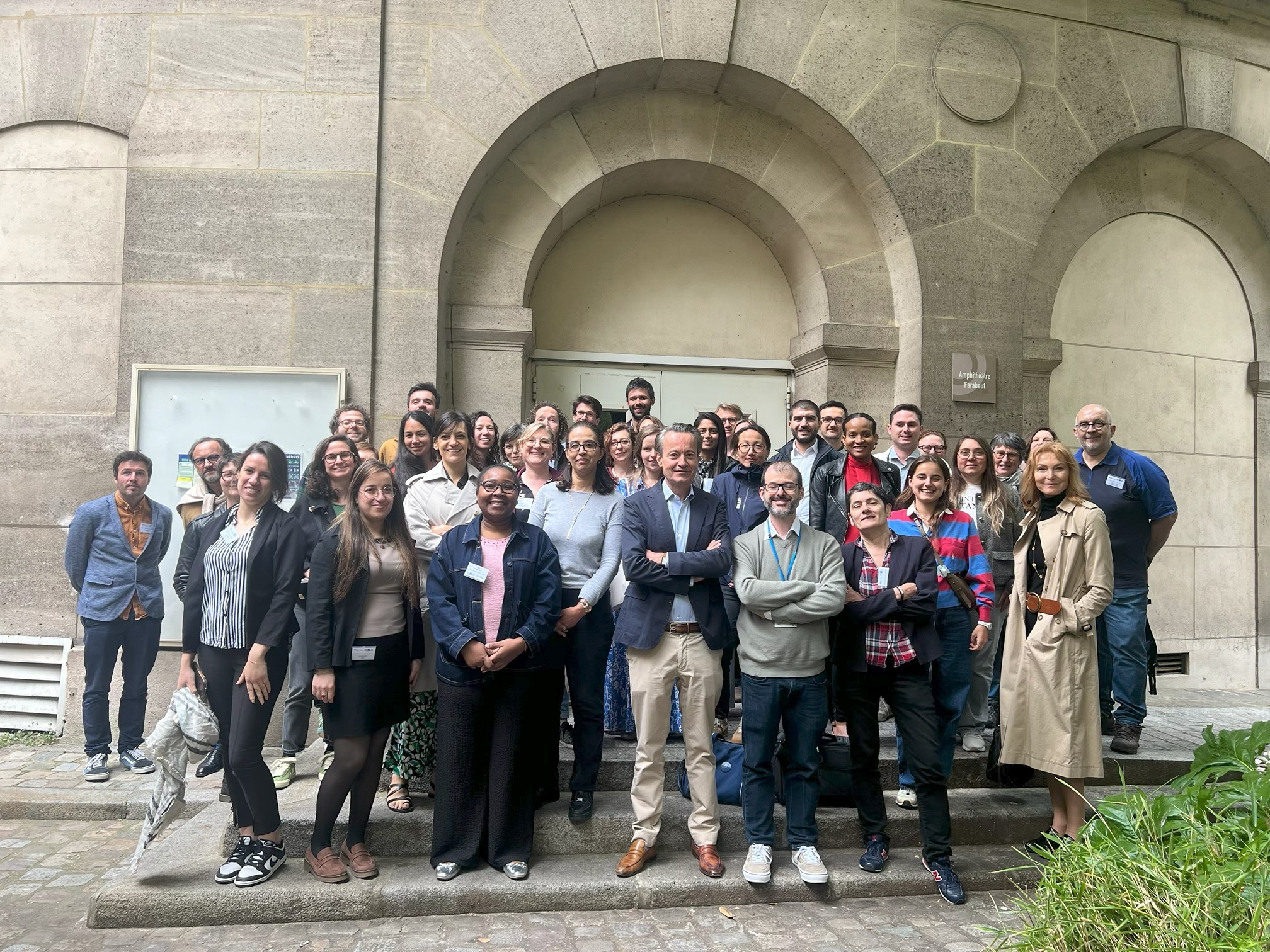
Le CRMR SPIN@ a organisé, le vendredi 5 juin 2026, sa 3ᵉ Journée scientifique à la Faculté de Médecine de Sorbonne Université, sur le site des Cordeliers à Paris. Cette journée d’échanges a réuni, dans une ambiance conviviale et chaleureuse, des professionnels de santé, des chercheurs ainsi que des représentants de l’association Spina Bifida et Handicaps Associés (ASBH). Le CRMR SPIN@ remercie chaleureusement l’ensemble des participants pour leur présence, la qualité des présentations et la richesse…
-

T(R)OUSS’AU SPORT 2026 : deux journées dédiées à l’inclusion par le sport à l’hôpital Trousseau AP-HP
Les 25 et 26 septembre 2026, l’hôpital Armand-Trousseau a célébré la cinquième édition de T(R)OUSS’AU SPORT. Cet événement porté par…
-

Voix de chercheur : Spina Bifida et chirurgie in utero par le Dr Guilbaud
À l’occasion de la semaine de la recherche clinique, du 18 au 22 mai 2026, l’AP-HP met à l’honneur pour…
-
4ᵉ journée « PRIUM » au Jardin des Plantes
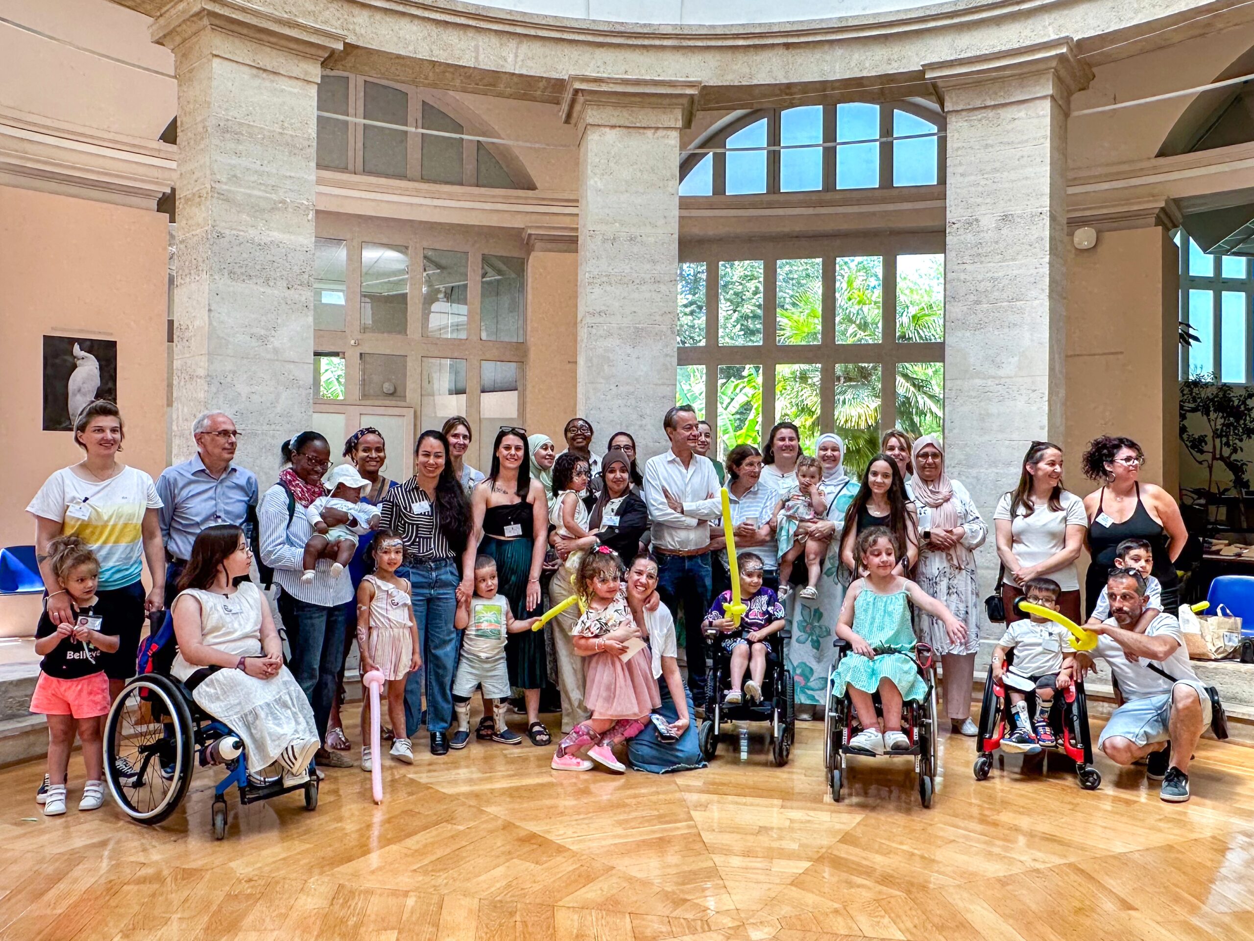
Le mercredi 17 juin 2026, le centre coordonnateur Spin@, en collaboration avec l’association de patients Spina Bifida et Handicaps Associés…
Nos publications
Brigato, Paolo; Palombi, Davide; Salvatore, Sergio De; Sessa, Sergio; de Saint Denis, Timothee; Oggiano, Leonardo; Gregori, Lorenzo Maria; Magistrelli, Andrea; Costici, Pier Francesco
Dans: Spine Deform, vol. 14, no. 4, p. 1121–1133, 2026, ISSN: 2212-1358.
@article{pmid41611975,

title = {Assessing pedicle morphology in pediatric spinal deformity: can magnetic resonance imaging match computed tomography? A systematic review},

author = {Paolo Brigato and Davide Palombi and Sergio De Salvatore and Sergio Sessa and Timothee de Saint Denis and Leonardo Oggiano and Lorenzo Maria Gregori and Andrea Magistrelli and Pier Francesco Costici},

doi = {10.1007/s43390-026-01290-5},

issn = {2212-1358},

year  = {2026},

date = {2026-07-01},

journal = {Spine Deform},

volume = {14},

number = {4},

pages = {1121--1133},

abstract = {BACKGROUND: Pediatric spinal deformities frequently require surgical correction, and precise preoperative planning is essential to ensure optimal outcomes and minimize complications. A key aspect of planning involves assessing pedicle morphology to determine appropriate screw sizing. This systematic review examines whether magnetic resonance imaging (MRI) can serve as a reliable, radiation-free alternative to computed tomography (CT). While MRI shows promising potential, current evidence presents mixed results regarding its accuracy and clinical applicability compared to CT.nnMETHODS: Following PRISMA guidelines, a systematic review was performed. Literature from Medline, Scopus, Embase, and the Cochrane Library, up to August 2025, was analyzed. Search terms included "scoliosis," "pediatric deformity," "pedicle morphology," "pedicle dimension," "magnetic resonance imaging," and "computed tomography." The extracted data included study details, patient demographics, scoliosis etiology, radiographic techniques, pedicle measurement methods, and outcomes of MRI-CT comparison. Risk of bias was assessed using the MINORS tool.nnRESULTS: Of 319 identified studies, 6 met the inclusion criteria (n = 428 patients). A total of 12,633 pedicles were analyzed across the studies, with MRI and CT used for preoperative assessment. MRI showed good concordance with CT for most measurements though accuracy decreased for more severe pedicle abnormalities. MRI was generally reliable for preoperative planning but had limitations, especially for pedicle sizing.nnCONCLUSION: While MRI offers a radiation-free alternative for preoperative assessment of pedicle morphology in pediatric spinal deformity surgery, current evidence is limited to retrospective studies and shows variable accuracy, particularly in severe or complex cases. CT generally remains more reliable for precise pedicle evaluation and continues to represent the standard of care. Future advancements in MRI technology may help improve its accuracy and expand its role in clinical practice, but stronger prospective evidence is needed before it can be considered a full substitute for CT.},

keywords = {},

pubstate = {published},

tppubtype = {article}

}
Parc, Enora; Jolivet, Eugénie; Maillet, Corentin; Athiel, Yoann; Jouannic, Jean-Marie; Guilbaud, Lucie
Dans: Prenat Diagn, vol. 46, no. 8, p. 1258–1266, 2026, ISSN: 1097-0223.
@article{pmid42141252,

title = {Impact of Fetoscopic Surgery on the Myometrium: Experimental Study in an Ovine Model of Myelomeningocele},

author = {Enora Parc and Eugénie Jolivet and Corentin Maillet and Yoann Athiel and Jean-Marie Jouannic and Lucie Guilbaud},

doi = {10.1002/pd.70177},

issn = {1097-0223},

year  = {2026},

date = {2026-07-01},

journal = {Prenat Diagn},

volume = {46},

number = {8},

pages = {1258--1266},

abstract = {OBJECTIVE: To investigate uterine healing at delivery after fetoscopic myelomeningocele (MMC) closure in an ovine model.nnMETHODS: This descriptive study was conducted in Romane ewes. Fetal MMC lesions were created after hysterotomy at 75 days of gestation. Fetoscopic MMC closure using three trocars was performed at 90 days of gestation and cesarean delivery at 143 days. Subtotal hysterectomy was performed, and each hysterectomy specimen was examined in fresh condition. The areas of interest and the surrounding intact tissue were then sampled en bloc and fixed in 4% buffered formaldehyde solution for 3-6 weeks. Macroscopic appearance of the scars was assessed in fresh condition. Microscopic examinations were conducted in fixed samples.nnRESULTS: The uteri of six ewes were studied. Macroscopic examination revealed identifiable fetoscopic scars without any scar dehiscence. There was no evidence of a difference in thickness between scarred and healthy adjacent myometrium, although a slightly lower proportion of muscle fibers was observed in scarred areas.nnCONCLUSIONS: Our study demonstrates that myometrial healing of trocar orifices is complete 53 days after fetoscopic MMC closure in an ovine model. Given the limitations of an ovine model, further studies are needed to determine the risk of uterine rupture and the optimal obstetric management of patients after fetoscopic MMC closure.},

keywords = {},

pubstate = {published},

tppubtype = {article}

}
Radmayr, Christian; Abrahamsson, Kate; Bogaert, Guy; Dellenmark-Blom, Michaela; Feitz, Wout; Goyal, Anju; Jouannic, Jean-Marie; Lammers, Rianne; Maternik, Michal; Mosiello, Giovanni; Roozen, Sylvia; Sillen, Ulla; Stein, Raimund; Tekgül, Serdar; ‘t Hoen, Lisette; Gontard, Alexander Von; Walle, Johan Vande; Yuan, Yuhong; Nijman, Rien J M
Dans: Eur Urol Open Sci, vol. 88, p. 39–53, 2026, ISSN: 2666-1683.
@article{pmid42088080,

title = {EAU-ESPU-ERN eUROGEN-ERN ITHACA-ERN ERKNet-IFSBH Guidelines on Spinal Dysraphism in Children and Adolescents: Summary of the Guideline},

author = {Christian Radmayr and Kate Abrahamsson and Guy Bogaert and Michaela Dellenmark-Blom and Wout Feitz and Anju Goyal and Jean-Marie Jouannic and Rianne Lammers and Michal Maternik and Giovanni Mosiello and Sylvia Roozen and Ulla Sillen and Raimund Stein and Serdar Tekgül and Lisette 't Hoen and Alexander Von Gontard and Johan Vande Walle and Yuhong Yuan and Rien J M Nijman},

doi = {10.1016/j.euros.2026.04.002},

issn = {2666-1683},

year  = {2026},

date = {2026-06-01},

journal = {Eur Urol Open Sci},

volume = {88},

pages = {39--53},

abstract = {BACKGROUND AND OBJECTIVE: Neurogenic bladder in children and adolescents presents complex challenges requiring comprehensive management strategies. Traditional guideline development often lacks input from diverse stakeholders, potentially overlooking crucial aspects of care. We present a novel approach to guideline development that integrates perspectives from pediatric urologists, nephrologists, psychiatrists, and patient representatives from the very beginning of the guideline development process. For this purpose, the European Association of Urology (EAU) Paediatric Urology Guidelines Panel worked together with European Reference Networks (eUrogen, ITHACA, ERKNet), the ESPU (European Society for Paediatric Urology), as well as the International Federation for Spina Bifida and Hydrocephalus (IFSBH), a professional organization with global coverage for people living with disabilities.nnMETHODS: A broad literature search was performed covering the timeframe from 2020 to 2024. Recommendations were developed and rated as strong or weak, based on the quality of the evidence, benefit/ harm ratio, and potential patient preferences.nnKEY FINDINGS AND LIMITATIONS: Spinal cord disorders have a profound impact on urinary, renal, and bowel functions, as well as on sexuality and fertility issues, necessitating lifelong management to preserve quality of life and prevent complications, such as urinary tract deterioration in individuals with spinal dysraphism. Timely diagnosis and appropriate multidisciplinary therapeutic management are critical, with comprehensive clinical evaluations-such as urodynamic studies-playing a central role. Treatment strategies should be individualized, incorporating a multidisciplinary approach that addresses not only medical and functional aspects but also considerations related to sexuality and fertility. Continuous monitoring and follow-up care underscore the significance of sustained management for patients with spinal dysraphism.nnCONCLUSIONS AND CLINICAL IMPLICATIONS: This summary of the 2025 EAU/ESPU/ERN eUrogen/ERN ITHACA/ERN ErkNet/IFSBH guideline provides updated guidance for evidence-based management of children and adolescents with spinal dysraphism.},

keywords = {},

pubstate = {published},

tppubtype = {article}

}
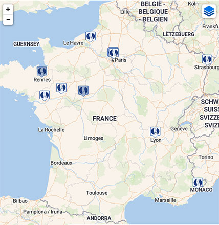
Carte interactive
Retrouvez l’ensemble des centres constituant le CRMR Spin@ sur notre carte géographique interactive